Picture 1 of 1
Stock photo

Picture 1 of 1
Stock photo
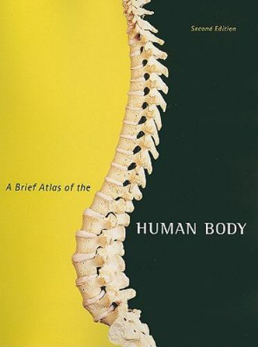
Brief Atlas of the Human Body by Matt Hutchinson, Elaine Marieb, Patricia Wilhelm and Jon Mallatt (2010, Spiral)
B
Brenham Book Company (1039)
93.7% positive feedback
Price:
$55.44
+ $4.25 shipping
Returns:
30 days returns. Buyer pays for return shipping. If you use an eBay shipping label, it will be deducted from your refund amount.
Condition:
This full-color atlas is packaged with every new copy of the text, and includes 107 bone and 47 cadaver photographs with easy-to-read labels. This edition of the atlas contains a comprehensive histology photomicrograph section featuring over 50 slides of basic tissue and organ systems. Featuring photos taken by renowned biomedical photographer Ralph Hutchings, this high-quality photographic atlas makes an excellent resource for the classroom and laboratory, and is referenced in appropriate figure legends throughout the text.
- Buy It NowBrief Atlas of the Human Body, A
Oops! Looks like we're having trouble connecting to our server.
Refresh your browser window to try again.
About this product
Product Identifiers
PublisherPearson Education
ISBN-10032166261X
ISBN-139780321662613
eBay Product ID (ePID)16038259034
Product Key Features
Number of Pages144 Pages
Publication NameBrief Atlas of the Human Body
LanguageEnglish
Publication Year2010
SubjectLife Sciences / Anatomy & Physiology (See Also Life Sciences / Human Anatomy & Physiology), Teaching Methods & Materials / Science & Technology, Atlases
FeaturesNew Edition
TypeTextbook
AuthorMatt Hutchinson, Elaine Marieb, Patricia Wilhelm, Jon Mallatt
Subject AreaScience, Education, Medical
FormatSpiral
Dimensions
Item Height0.4 in
Item Weight15.1 Oz
Item Length10.5 in
Item Width7.9 in
Additional Product Features
Edition Number2
Intended AudienceCollege Audience
TitleLeadingA
Dewey Edition22
Dewey Decimal612
Edition DescriptionNew Edition
SynopsisThis full-color atlas includes 107 bone and 47 cadaver photographs with easy-to-read labels. This edition of the atlas contains a comprehensive histology photomicrograph section featuring over 50 slides of basic tissue and organ systems. Featuring photos taken by renowned biomedical photographer Ralph Hutchings, this high-quality photographic atlas makes an excellent resource for the classroom and laboratory, and is referenced in appropriate figure legends throughout the text., This full-color atlas is packaged with every new copy of the text, and includes 107 bone and 47 cadaver photographs with easy-to-read labels. This edition of the atlas contains a comprehensive histology photomicrograph section featuring over 50 slides of basic tissue and organ systems. Featuring photos taken by renowned biomedical photographer Ralph Hutchings, this high-quality photographic atlas makes an excellent resource for the classroom and laboratory, and is referenced in appropriate figure legends throughout the text.
All listings for this product


Be the first to write a review


